Mast Cell Tumors on a Labrador
Finding a new lump or bump on your beloved Labrador can send any dog owner into a tailspin of worry. It's a terrifying moment, especially when you know that while many lumps are harmless fatty growths, some can be far more serious. For Labradors, one type of cancerous growth that often causes concern and requires careful attention is the mast cell tumor. This guide aims to thoroughly demystify mast cell tumors, helping you understand what they are, how to recognize them, and the steps involved in their diagnosis and treatment, so you can be prepared if this unwelcome visitor appears on your furry friend.
Understanding Mast Cell Tumors in Labradors
When you discover an unexpected lump on your Labrador, your mind immediately jumps to the worst-case scenario. It's perfectly natural to feel that panic, especially with a breed known for its robust health but also, sadly, a propensity for certain cancers. Mast cell tumors are one of those cancers, and understanding them is the first step in addressing the fear and uncertainty they bring.
What are Mast Cells and Their Role?
Let's start with the basics. Mast cells are a normal and vital part of your dog's immune system. Think of them as tiny alarm bells scattered throughout the body, particularly in areas like the skin, respiratory tract, and gastrointestinal tract. Their primary job is to respond to inflammation and allergic reactions. When triggered, these cells release various chemicals, including histamine, which causes local swelling, itching, and redness – the classic signs of an allergic reaction or inflammation. This is a good thing when your dog is fighting off a parasite or responding to a bee sting.
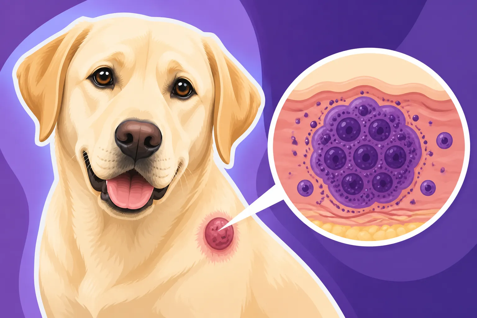
However, a mast cell tumor is essentially an uncontrolled proliferation of these mast cells. Instead of acting as helpful immune system components, they become cancerous, multiplying without regulation and potentially releasing their chemical contents indiscriminately. This uncontrolled release of histamine and other substances can lead to not only the visible tumor but also a host of systemic problems, which we'll delve into shortly. These tumors can be sneaky, sometimes looking like a benign wart or an insect bite, making early detection and veterinary consultation even more critical.
Why Labradors are Prone to Mast Cell Tumors
It’s an unfortunate reality that certain breeds seem to have a higher incidence of specific health issues, and Labradors are no exception when it comes to tumors. While breeds like Boxers, Boston Terriers, and Bulldogs are famously prone to mast cell tumors, Labradors also feature prominently on the list of breeds at elevated risk. This predisposition is thought to have a genetic component, though the exact genes and pathways are still being researched. It’s not about poor breeding; it’s simply a genetic lottery that sometimes doesn't go in their favor.
Mast cell tumors can appear virtually anywhere on a Labrador's body, both externally and internally. However, they are most commonly found on the skin, often on the trunk and limbs. They can also arise in internal organs such as the spleen, liver, lymph nodes, or bone marrow, which are much harder to spot without diagnostic imaging. The age at which they appear can vary widely, but they are more frequently diagnosed in middle-aged to older Labradors, though even younger dogs are not entirely immune. Recognizing that your Labrador might be more susceptible means being extra vigilant about any new lumps or bumps you find during your regular petting sessions. This awareness is crucial for early intervention and better outcomes.
Recognizing the Signs of a Mast Cell Tumor
The sheer variety in how mast cell tumors present themselves is what makes them so tricky and often a source of significant anxiety for Labrador owners. It's not always a clear-cut, textbook growth, and what might seem like an innocuous bump could be something far more concerning.
The 'Lump' That Changes
Many Labrador owners first notice a mast cell tumor as a single, solitary mass on or just under the skin. The appearance of these lumps is incredibly diverse, which is why it’s so important never to assume a lump is benign without veterinary evaluation. They can be:
- Firm or soft: Some feel like a hard knot, while others are squishy.
- Raised or flat: They might stand out like a wart or be subtle, just beneath the surface.
- Hairless or covered in fur: There's no consistent rule here.
- Reddish or skin-colored: Their color can vary, sometimes appearing inflamed.
- Slow-growing or rapid: Some seem to pop up overnight, others grow imperceptibly over weeks or months.
What's particularly concerning and a classic indicator of a mast cell tumor is a phenomenon often called "Darier's sign" or the "itch, switch, and swell" reaction. This means the tumor might suddenly become red, swollen, and itchy, often after being scratched, rubbed, or even just touched. This reaction is due to the unstable mast cells releasing their inflammatory chemicals. You might notice your Labrador licking or chewing at the spot more than usual, only for the lump to inflame and then subside. This cyclical change makes owners sometimes dismiss it as an allergic reaction or an insect bite. However, any lump that changes in size, shape, or color, or seems to come and go, must be investigated by a veterinarian immediately. It’s a key Warning Signs of Cancer in a Labrador that should never be ignored. Contrast this with Lipomas and Fatty Lumps on a Labrador, which typically remain soft, movable, and don't usually change size dramatically or become inflamed.
Systemic Symptoms to Watch For
While a skin lump is the most common manifestation, mast cell tumors are not always confined to the surface. As mentioned, they can arise internally or spread from a skin tumor to other parts of the body. When this happens, or when the tumor is particularly aggressive (high-grade), the constant release of histamine and other chemicals can wreak havoc throughout your Labrador's system, leading to what are known as paraneoplastic syndromes.
These systemic symptoms can be vague and easily mistaken for other illnesses, making diagnosis even harder:
- Gastrointestinal issues: Vomiting, diarrhea, and loss of appetite are common because histamine stimulates gastric acid production, which can lead to stomach ulcers. You might see black, tarry stools (melena) if an ulcer is bleeding.
- Lethargy and weakness: Your normally energetic Labrador might seem unusually tired, less interested in walks, or just generally "off."
- Weight loss: Despite a healthy appetite (or sometimes with a decreased appetite), your dog might start to lose weight.
- Anemia: Chronic blood loss from gastrointestinal ulcers can lead to anemia, causing pale gums and weakness.
- Swollen lymph nodes: If the cancer has spread to regional lymph nodes, they might feel enlarged.
If your Labrador is experiencing any of these generalized symptoms, especially in conjunction with a suspicious lump on their skin, it's crucial to seek veterinary attention promptly. These signs indicate that the tumor might be more aggressive or has potentially spread, requiring a comprehensive diagnostic workup beyond just the visible lump. Early intervention truly makes a difference in these cases.
The Path to Diagnosis and Staging Mast Cell Tumors
The moment you notice a lump, your vet becomes your most important ally. A thorough diagnostic process is critical to identify mast cell tumors, determine their aggressiveness, and check for any spread, all of which will inform the best treatment plan for your Labrador.
Initial Vet Visit and Fine Needle Aspirate (FNA)
When you first bring your Labrador in with a new lump, your veterinarian will perform a thorough physical examination, noting the size, location, consistency, and any other characteristics of the mass. This is where the detective work begins. The next crucial step is usually a Fine Needle Aspirate (FNA).
During an FNA, your vet will use a very thin needle (similar to those used for vaccinations) to collect a small sample of cells directly from the lump. This is a quick procedure, often done in the exam room without sedation, though a highly anxious dog might need light calming. The collected cells are then smeared onto a slide, stained, and examined under a microscope. This is where mast cell tumors often give themselves away: mast cells have distinctive purple granules that are easily visible to a trained eye.
An FNA is incredibly valuable because it's minimally invasive and can often provide an immediate preliminary diagnosis. If mast cells are identified, it confirms suspicion. However, an FNA also has limitations. It only samples a small portion of the tumor, and sometimes, a definitive diagnosis or an accurate grading (how aggressive the tumor is) can't be made from an FNA alone. The sample might not be representative, or the cells might not be clearly defined. If the FNA is inconclusive, or if mast cells are found but more information is needed, your vet will recommend a biopsy.
Biopsy and Histopathological Grading
While an FNA can suggest a mast cell tumor, a surgical biopsy is the gold standard for a definitive diagnosis and, most importantly, for grading the tumor. This involves surgically removing a piece of the tumor (incisional biopsy) or, ideally, the entire tumor with surrounding healthy tissue (excisional biopsy). The tissue sample is then sent to a veterinary pathologist who specializes in examining tissues and diagnosing diseases.
The pathologist will analyze the tissue under a microscope to confirm the presence of mast cells and, critically, assign a grade to the tumor. This grading system is paramount as it directly correlates with the tumor's biological behavior and metastatic potential (its likelihood to spread). The most commonly used system categorizes mast cell tumors into:
- Grade I (Low Grade): These tumors are well-differentiated, meaning their cells look fairly normal. They are generally considered benign or locally aggressive and have a low chance of spreading.
- Grade II (Intermediate Grade): This is the most common grade. These tumors have some abnormal cellular characteristics and are moderately aggressive. They have a variable potential to spread and recur, making their management more complex.
- Grade III (High Grade): These are poorly differentiated and highly aggressive tumors. Their cells look very abnormal, and they have a high metastatic potential, meaning they are much more likely to spread quickly to other organs.
There's also a newer two-tier system that classifies mast cell tumors as either low-grade or high-grade, which many oncologists find more clinically useful. Regardless of the system used, obtaining a grade is crucial for predicting prognosis and guiding treatment decisions.
Staging for Spread
Once a mast cell tumor has been diagnosed and graded, the next critical step is 'staging.' Staging determines if the cancer has spread from its original site to other parts of your Labrador's body. This is especially important for higher-grade tumors, but even lower-grade tumors can sometimes surprise us. The process of staging often involves several diagnostic tests:
- Blood work: A complete blood count (CBC) and biochemistry panel can assess overall organ function and look for signs of systemic illness related to the tumor.
- Urinalysis: To check kidney function and overall health.
- Abdominal ultrasound: This allows the vet to visualize internal organs like the spleen, liver, and abdominal lymph nodes, which are common sites for mast cell tumor metastasis. If any abnormalities are seen, further FNAs or biopsies might be recommended.
- Chest radiographs (X-rays): To check for any signs of spread to the lungs, though mast cell tumors rarely spread directly to the lungs, making this less common than abdominal checks.
- Lymph node aspiration: If nearby lymph nodes feel enlarged or suspicious, an FNA of these nodes is often performed to check for cancer cells.
- Bone marrow aspiration: In very aggressive cases or when other tests indicate widespread disease, a bone marrow biopsy might be considered.
This comprehensive staging process provides a full picture of the cancer's extent, allowing your veterinary oncologist to create the most effective, tailored treatment plan for your Labrador. Without staging, you might treat the visible tumor only to find out later that the cancer had already spread internally, significantly impacting prognosis.
Navigating Treatment Options for Mast Cell Tumors
Receiving a cancer diagnosis for your Labrador is heartbreaking, but it's important to remember that mast cell tumors, especially lower-grade ones detected early, are often treatable. The specific treatment approach will depend heavily on the tumor's grade, stage, and location.
Surgical Excision as the Primary Tool
For most localized mast cell tumors, surgery is the cornerstone of treatment. The goal of surgery is to remove the entire tumor with wide, clean margins, meaning a significant amount of healthy tissue surrounding the visible tumor is also removed. This is crucial because mast cells can infiltrate surrounding tissues in ways that aren't visible to the naked eye. The saying in veterinary oncology is "the first surgery is the best chance for a cure."
The size and location of the tumor can greatly influence the success of surgical excision. A small tumor on the trunk might be relatively straightforward to remove with wide margins. However, a tumor on a limb, digit, or the face presents a much greater challenge due to the limited amount of excess skin available for removal and closure. If the margins are found to be "dirty" or "incomplete" after pathology review (meaning cancer cells are still present at the edges of the removed tissue), further treatment options will need to be considered. This might include a second surgery, radiation therapy, or close monitoring, depending on the tumor's grade and the perceived risk.
Recovery from surgery typically involves pain management and activity restriction, similar to other surgical procedures. Your vet will provide detailed post-operative care instructions, and it's vital to follow them closely to ensure proper healing.
Radiation Therapy and Chemotherapy
When surgery alone isn't sufficient or possible, or when the tumor is more aggressive, other therapies come into play.
Radiation Therapy: This treatment uses high-energy radiation to kill cancer cells or shrink tumors. It's often recommended in situations where:
- Incomplete surgical margins: If the pathologist confirms that cancer cells were left behind after surgery, radiation can target these microscopic cells.
- Non-resectable tumors: Tumors that are too large or in difficult locations (e.g., around the eye, on a digit) to be completely removed surgically.
- Local recurrence: If a tumor reappears in the same area after initial surgery.
Radiation therapy typically involves multiple short sessions over several weeks, requiring the dog to be sedated or anesthetized for each treatment. This can be a significant commitment in terms of time and cost. It's important to discuss the potential side effects with your veterinary oncologist, which can include skin irritation and hair loss in the treated area.
Chemotherapy: This involves using drugs to kill cancer cells throughout the body. It's usually reserved for:
- High-grade tumors: Grade II (with concerning features) or Grade III mast cell tumors, due to their higher risk of metastasis.
- Metastatic disease: When the cancer has already spread to other organs or lymph nodes.
- Tumors that cannot be surgically removed or radiated: As a palliative measure or to reduce tumor size before other treatments.
There are several chemotherapy protocols for mast cell tumors. Common medications include prednisone (a steroid often used alone or with other drugs), vinblastine, lomustine, and newer targeted therapies like toceranib (Palladia) or masitinib (Kinavet). These targeted therapies work by inhibiting specific pathways involved in cancer growth. The choice of chemotherapy depends on the tumor's grade, stage, and your Labrador's overall health. Chemotherapy for dogs is generally better tolerated than in humans, with fewer severe side effects, but careful monitoring by your vet is essential. Understanding Chemotherapy Costs for a Labrador Retriever is an important part of making informed decisions for your pet's care.
Palliative and Supportive Care
For Labradors with advanced, widespread, or high-grade mast cell tumors where a cure is unlikely, the focus shifts to palliative care. This approach aims to maximize your dog's quality of life and minimize discomfort rather than aggressively pursuing a cure.
Palliative care strategies include:
- Pain management: Using medications to control pain associated with the tumor or its effects. Learning to recognize Reading Pain in a Stoic Labrador is crucial here.
- Antihistamines: Drugs like diphenhydramine (Benadryl) or cimetidine can help counteract the effects of histamine released by mast cells, reducing itching, swelling, and stomach upset.
- Gastroprotectants: Medications like omeprazole or famotidine can help prevent or treat stomach ulcers caused by excessive histamine.
- Steroids: Prednisone can sometimes shrink mast cell tumors, even in advanced stages, and improve appetite and overall well-being.
- Nutritional support: Ensuring your Labrador maintains a healthy weight and receives adequate nutrition.
This type of care is deeply personal and focuses on making every day count. It involves open communication with your veterinary team to ensure your Labrador is as comfortable and happy as possible for as long as possible. Sometimes, the kindest decision is to focus on comfort rather than aggressive treatments that may cause more suffering.
Prognosis and Living with a Labrador with an MCT
Navigating a cancer diagnosis for your Labrador is an emotional rollercoaster, and understanding the potential outcomes and ongoing management is crucial for preparing yourself and providing the best care.
Factors Influencing Outcome
The prognosis for a Labrador with a mast cell tumor is highly variable and depends on several key factors:
- Tumor Grade: This is arguably the most significant prognostic indicator. Low-grade (Grade I) tumors, especially if completely removed, often have an excellent prognosis with long survival times. High-grade (Grade III) tumors carry a guarded to poor prognosis due to their aggressive nature and high metastatic potential. Intermediate-grade (Grade II) tumors have a more variable prognosis, often requiring further assessment of specific features like mitotic index (rate of cell division).
- Tumor Stage: Whether the tumor has spread (metastasis) is another critical factor. A tumor that has spread to lymph nodes or internal organs has a much worse prognosis than a localized tumor.
- Location of the Tumor: Certain locations are associated with a worse prognosis, regardless of grade. These "aggressive sites" include tumors on the muzzle, digits, mucous membranes (like the mouth or anus), or internal organs. These sites are often difficult to get wide surgical margins, increasing the risk of recurrence.
- Completeness of Surgical Margins: If the surgical margins are clean (no cancer cells at the edges), the prognosis is generally better. Incomplete margins often necessitate further treatment.
- Presence of Systemic Signs: If your Labrador is showing signs of histamine release (e.g., gastrointestinal issues), this can complicate treatment and indicate a more aggressive tumor.
- Breed: While Labradors are generally prone, their prognosis is often considered more favorable than, say, Boxers for a given grade.
- Recurrence: A tumor that has recurred after initial treatment often carries a more guarded prognosis.
Your veterinary oncologist will combine all these factors to give you the most accurate prognosis for your individual Labrador, but remember, every dog is an individual, and outcomes can sometimes defy expectations.
Life After Diagnosis: Monitoring and Support
Even after successful treatment, vigilance remains key when living with a Labrador who has had a mast cell tumor. Regular follow-up appointments with your veterinarian or oncologist will be scheduled to monitor for any signs of recurrence at the original site or the development of new tumors. Labradors prone to one mast cell tumor are often predisposed to developing others in different locations.
During these check-ups, your vet will perform a thorough physical exam, palpating lymph nodes and checking for any new lumps or bumps. Depending on the original tumor's grade and stage, periodic blood work or imaging studies might also be recommended.
As an owner, you become your Labrador's first line of defense. Continue to perform regular "lump checks" when you're petting or grooming your dog. Pay attention to any changes in their behavior, appetite, or energy levels that could hint at systemic issues. Maintaining a good quality of life is paramount. This includes a balanced diet, appropriate exercise (within your vet's recommendations, especially after surgery), and plenty of love and comfort. Remember that your Labrador relies on you, and your emotional well-being also matters in this journey.
Financial Considerations and Support
Treating cancer in any pet can be an unexpected and significant financial burden. Costs can range from hundreds for a simple surgical removal of a low-grade tumor to thousands or even tens of thousands of dollars for complex surgeries, radiation, or extended chemotherapy protocols. This can be a huge shock, and it’s a valid concern for any pet parent.
It's important to have an open conversation with your veterinary team about the costs associated with each diagnostic test and treatment option. They can often provide estimates and help you prioritize. This is where options like pet insurance can truly be a lifesaver. Many pet owners find that a good insurance policy helps them make medical decisions based on what's best for their pet, not just what they can afford out-of-pocket. Providers like Healthy Paws often cover a significant portion of cancer treatments, including diagnostics, surgery, chemotherapy, and even some alternative therapies, though it's crucial to understand policy details regarding deductibles, reimbursement rates, and waiting periods. For instance, Whether Insurance Covers Labrador Hip Dysplasia is a common question, and similarly, you should understand how cancer is covered. If you don't have insurance, consider setting up How Big an Emergency Fund a Labrador Needs or looking into payment plans or financial aid organizations. Unfortunately, Pre-Existing Conditions on a Labrador Policy are usually not covered, so getting insurance before a diagnosis is key.
Remember, you're not alone in this. Your veterinary team is there to support you through every decision, helping you weigh the options to give your Labrador the best possible care while also considering your family's circumstances.
Disclaimer
The information provided in this article is for informational purposes only and does not constitute medical advice. Always consult with a qualified veterinarian for any health concerns or before making any decisions about your pet's medical care.